-
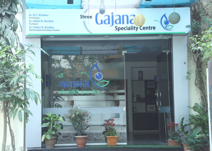
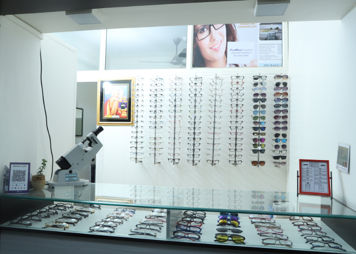
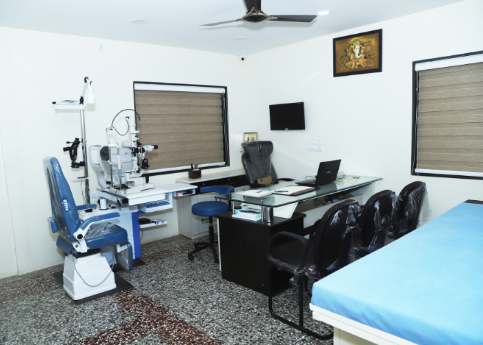
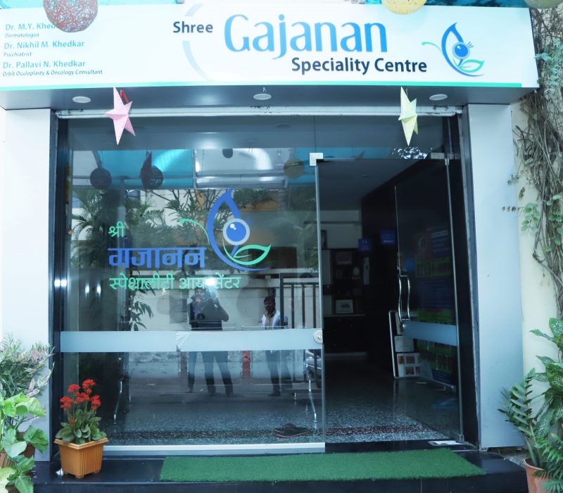
 Shree Gajanan SpecialityEyeCenterOne of the best ophthalmic plastic surgery
center. We provide treatment for eyelid &
orbit diseases and trauma-related problems. -
 Ocular Prosthesis/Artificial EyesAn artificial eye implanted in people who have
lost their eyes due to a variety of reasons.
The implantation of the artificial eyes helps
in the restoration of patients' confidence by
enhancing their physical appearance. -
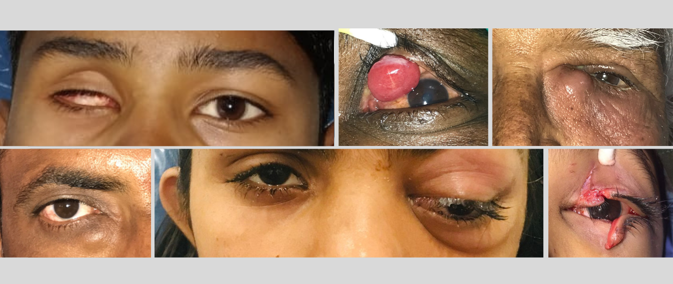 We are ProvidingTreatments For- Eye Check-Up - Droopy Eyelids/Ptosis - Eye Cancer - Facial Spasms & Botox - Watering Eyes-Dacryology - Socket Treatment & Artificial Eye
About
Shree Gajanan Eye Speciality Centre
We Protect Your Vision
Shree Gajanan Hospital is a high-end multispecialty eye clinic in Aurangabad, Maharashtra, that offers a wide range of treatments for Eyelid and orbit disorders and trauma, Top eye plasty services. We are also known as Aurangabad top eye clinic. We provide professional eye care services such as comprehensive diagnosis and clinical/surgical management of functional and aesthetic abnormalities in the eyelid, the orbit region (bony structure around the eye), and the lacrimal system (tear drainage system). Dr. Pallavi Khedkar, our oculoplastic surgeon, is experienced in diagnosing patients’ eye disorders and repairing them with greater care. Dr. Pallavi Khedkar is a leading Eye plastic surgeon in Aurangabad. We have a long history of delivering individualized care and dealing with the most difficult cases. Our team of oculoplastic surgeons, faciomaxillary surgeons & anesthetists, and staff work together as a team to ensure that your needs are met. To consult the best eye doctor, go to Shree Gajanan Hospital.
Facilities Available
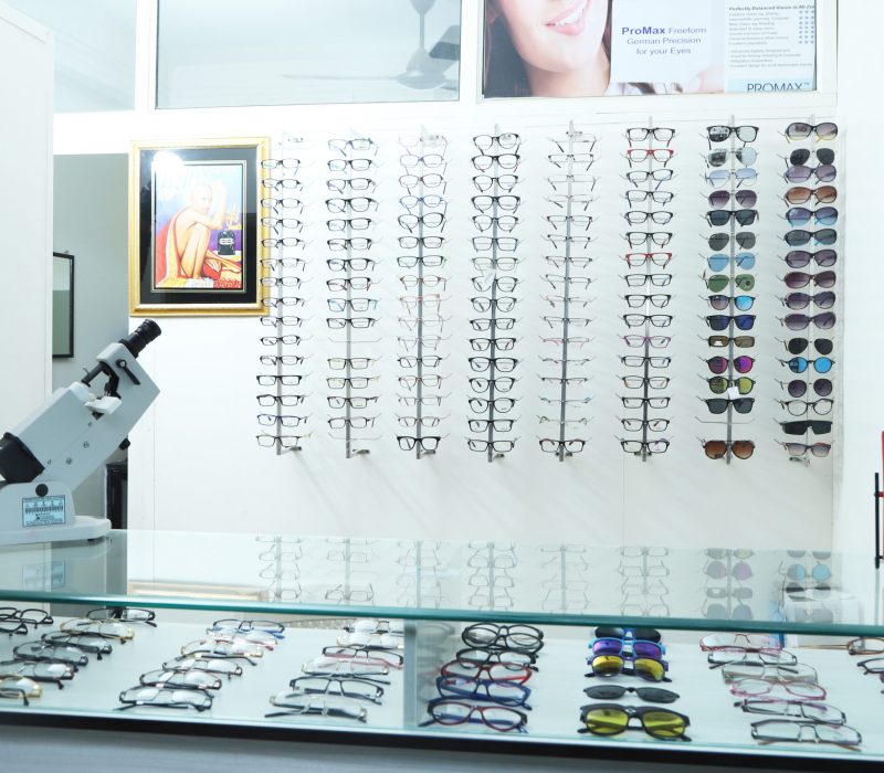
- Assured Diagnosis Of Lid & Periocular Disease
- Complete treatment Of Lid Diseases
- Eye Cancer
- Treatment Of Congenital Eye Anomalies
- Botox Clinic
- Ptosis Clinic
- Ocular Prosthesis Lab
- Dacryology Clinic
- Orbital Trauma Center
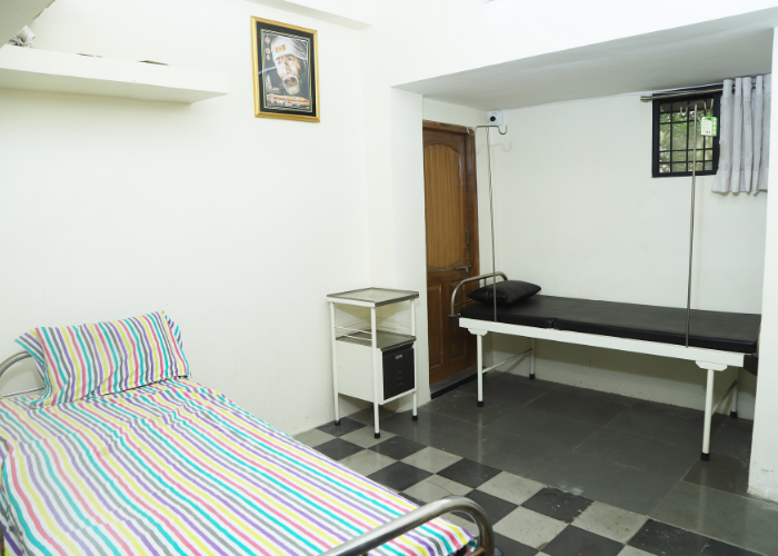
- Well Equipped Operation Theater
What is Oculoplastic Surgery?
The specialty of oculoplastic surgery deals with the eyelids, the bony orbit around the eye, the eyebrows, and the tear ducts and glands. Dr. Palllavi Khedkar is a trained Eye Plastic Surgeon in Aurangabad with a particular interest in performing facial cosmetic procedures.
When You Need to Visit Oculoplasty Clinic
- Eyelid deformity (Ptosis, Coloboma)
- Turning the edges of the eyelid in or out(Entropion/Ectropion)
- Painful eye or congenital malformed eye.
- Persistent watery eyes.
- Mass on the eyelid (Benign/Malignant)
- The eye may feel large or out of the groove.
- Fractures of the orbital bones due to accident.
Why Choose An Oculoplastic Surgeon?
The eyelids and surrounding structures are complex and vital structures that are critical to vision preservation. Furthermore, the eyes are the first feature on a person’s face that everyone sees. Oculoplastic surgeons are highly qualified to conduct complex eyelid, orbital, and cosmetic surgery around the eye, as well as provide care for the eye itself, since they are skilled in both ophthalmology and oculoplastic surgery.
An oculoplastic surgeon usually completes a three-year residency in ophthalmology (diseases of the eye). Following that, they complete hands-on training in oculoplastic surgery. They are trained in the treatment and surgery of all eyelid and orbital disorders, trauma, tumors, as well as aesthetic surgery and cosmetic procedures involving the eye and face. As a result, oculoplastic surgeons like Dr. Pallavi Khedkar have an unrivaled level of preparation, skills, and experience to tackle any kind of complex plastic surgery problem that arises.
About Dr. Khedkar
Dr. Pallavi Khedkar- Eye Plastic Surgeon in Aurangabad
M.B.B.S. ( J. J. Hospital, Mumbai )
D.N.B. ( Sankara Nethralaya, Chennai)
F.A.E.H. ( Aravind Eye Hospital, Madurai)
Orbit Oculoplasty Surgeon.
Consultant Ocular Oncologist.
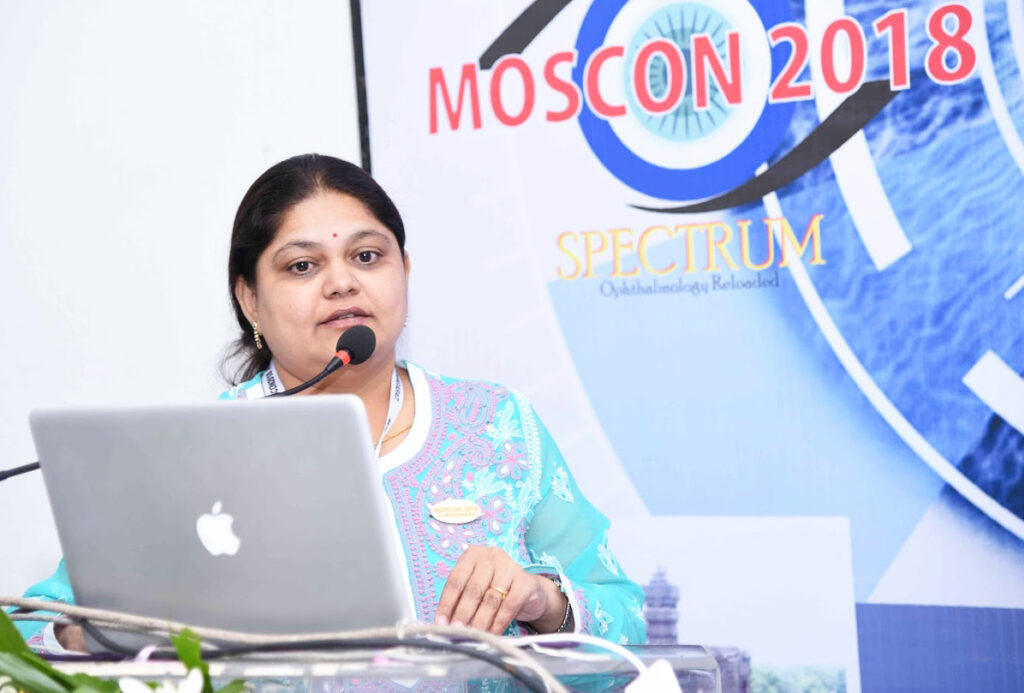
Dr. Pallavi Khedkar is the only Ophthalmic plastic surgeon & orbit consultant in Aurangabad District. She is known as a leading Eye Plastic Surgeon in Aurangabad. She has graduated from grant medical college, J. J. Hospital Mumbai. She achieved (DNB) from the world-renowned center in ophthalmology Sankara Nethralaya Chennai. She accomplished a long-term fellowship in oculoplastic, orbital disorders, and ocular oncology from the internationally renowned institute Aravind Eye Hospital, Madurai. She was an active team member in Arvind Hospital’s Ocular Oncology Unit. She has presented papers in various national conferences held at Bhopal, Delhi, Kolkata, Pune, Mumbai, Kodaikanal, etc.
Eye Check-Up
Eye Check-Up
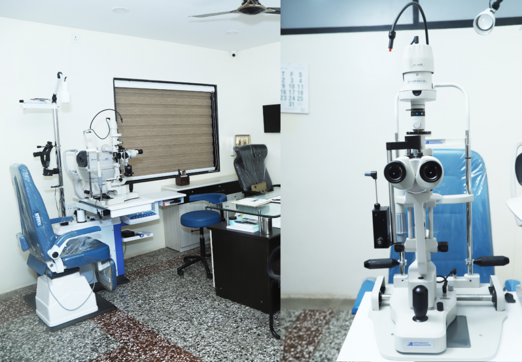
A regular eye exam is essential for clear eyesight and prevents complications or progress of ocular problems.
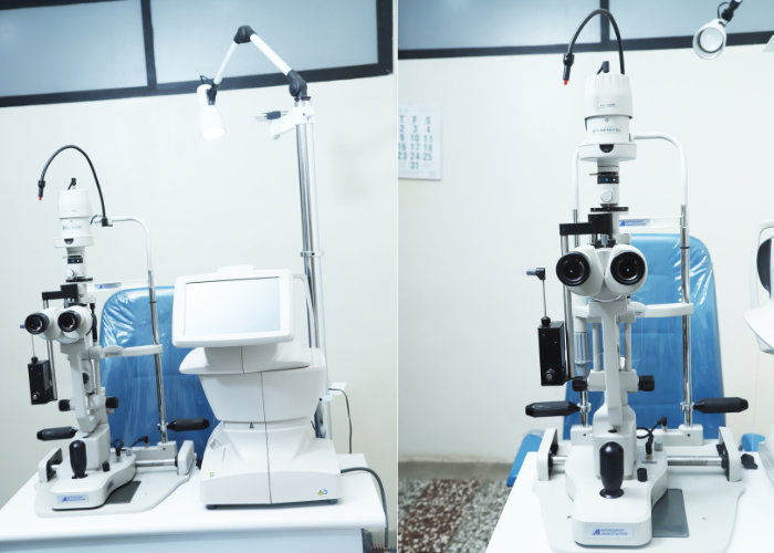
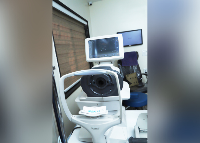
Routine eye exams for cataracts, visual acuity testing, tonometry, slit lamp exam, dilated eye exam with indirect ophthalmoscopy.
Evaluation of specific eyelid diseases and lacrimal drainage problems is done along with few diagnostic tests.
Eyelid Diseases
MALPOSITION OF THE EYELID MARGINS
Entropion
In entropion, the eyelid margin turns in. This causes the eyelashes to rub on the eye itself.
This leads to redness, watering, and foreign body sensation in the eye.
Who can have it?
Commonly it is seen in older age groups generally after 60years,
It can be present at birth or acquired after trauma, tumors, inflammation, or can occur with old age (most common).
Symptoms
Patients suffer from red eyes, pain, infections, and tearing. The symptoms are typically more severe and disabling than Ectropion.
Treatment
Surgical correction of the lid margin position Usually, incisions are cosmetically oriented Results of surgery are very reliable and have the course of 2 weeks of healing
Please Note: Trimming of eyelashes can be counterproductive and cause larger thicker lashes which can further damage the surface of the eye.

Ectropion
Causes
It can be present at birth or acquired after trauma, tumors, or inflammation on the skin or can occur with old age.
Symptoms
This leads to a variety of symptoms including eyeball irritation, a red-eye, watering , pain, infections.
Treatment
Mainly surgical correction is needed
Sometimes skin grafts and multiple injections around the scar are advisable
Please note: Malposition of the eyelid margins results in eye irritation and watering from the eye.

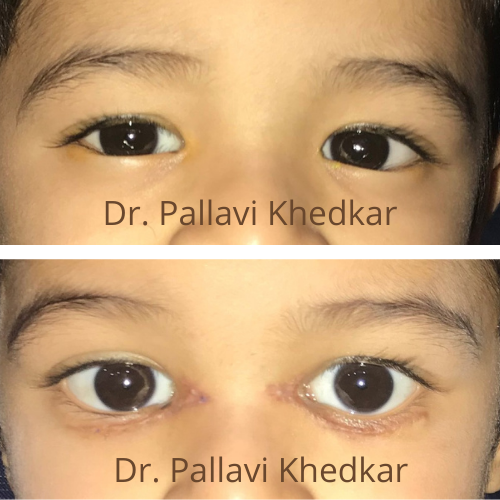
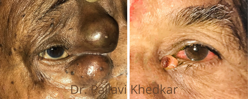
Congenital Entropion
Case 1
A boy 4 yrs old presented with congenital entropion with medial epicanthal folds. He presented with watering and redness. He underwent surgical correction of lower lid entropion and epicanthal folds under general anesthesia. He recovered well within 1 week
Tumours Eye & Adnexa
Ocular Oncology
Ocular oncology is a niche area of ophthalmology with only a handful of experts across the country. Dr. Pallavi Khedkar is trained in the diagnosis and management of ocular and orbital tumors in Aravind eye hospitals Madurai and has worked as a team with experts in this field. Ocular Oncology encompasses all tumors – benign and malignant that can affect every part of the eye – the eyelid, conjunctiva, choroid, retina, optic nerves, and the orbit as well.
Tumors or inflammation can occur behind the eye and often push the eye forward causing a prominent and protruded eye (proptosis) and include the following causes.
Causes
- Dermoid
- Lymphoma
- Lacrimal gland tumors
- Vascular tumors (hemangioma or lymphangioma)
- Optic nerve tumors
The patient may have dimness of vision, double vision, and cosmetic concerns due to bulging eyes, red eyes, watery and painful eyes.
Orbitotomy with subsequent treatment. Whenever possible, orbital tumors need to be totally excised and sent for biopsy.
Some tumors may need chemotherapy or radiotherapy for adjuvant treatment. In some advanced tumors, it may be necessary to proceed with the removal of eye & orbital contents (enucleation).
After a thorough examination and CT imaging, the concern can be addressed to cure and through a decent cosmetic approach leaving minimal to no after surgery visible scars
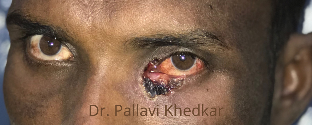
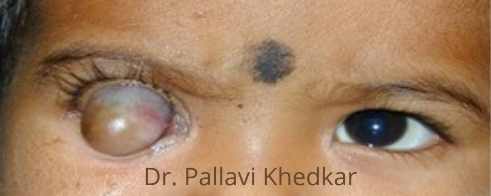
A few representative cases of ocular oncology which are treated and managed by Dr. Pallavi Khedkar are shown here.
Case 1
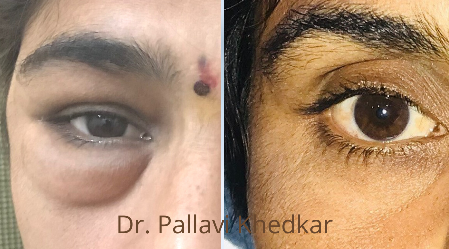
A 60-year-old lady presented with a history of a slow-growing ulcerated mass just below the lower eyelid. The lesion was treated with excision and a skin graft was placed. The lesion was a basal cell carcinoma confirmed by histopathology.
Case 2
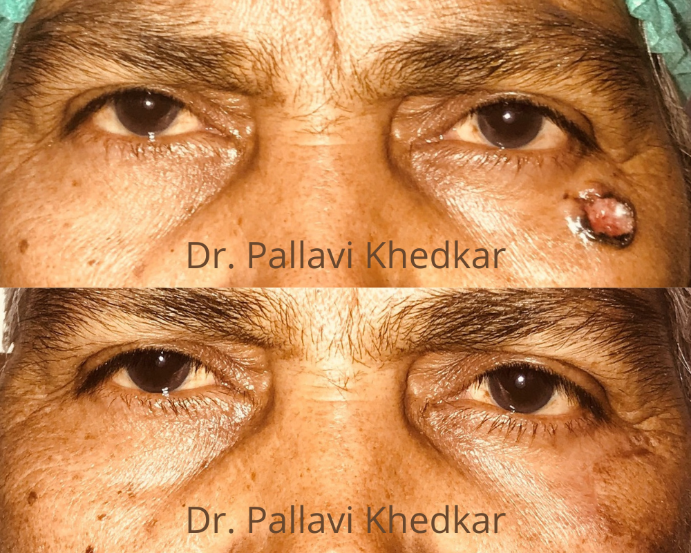
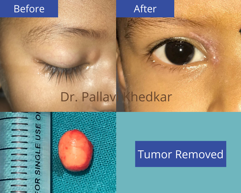
A 2-year-old child presented with a mass around the eye progressively growing in size. He underwent excision biopsy with a cosmetically acceptable scar and cystic solid mass was sent for histopathology. Mass was confirmed to be dermoid cyst – benign mass.
Orbital Tumor
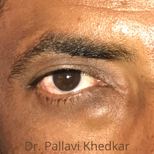
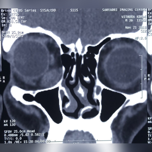
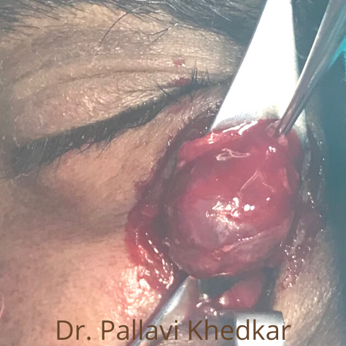
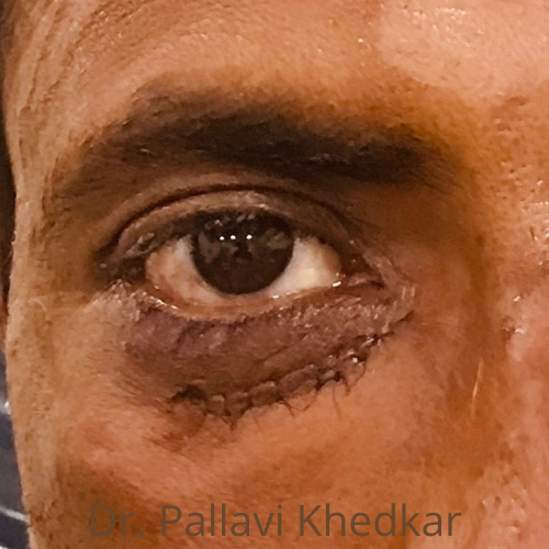
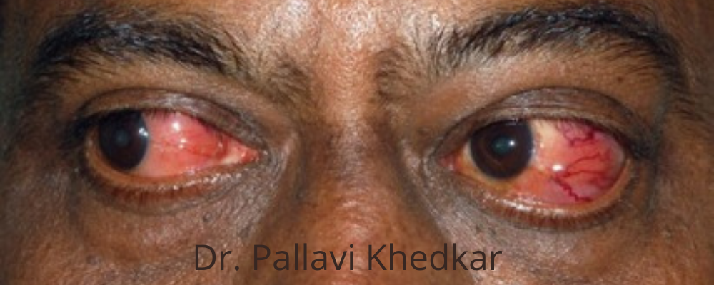
Thyroid Eye Disorder
Patients with thyroid abnormalities often manifest eyelid and orbital disease. Often the upper and lower lids retract and the eyes become prominent (proptosis). The pathophysiology behind the symptoms and signs is the autoimmune-mediated deposition of protein and swelling of muscles of the eyeball.
Swollen eyes, redness, protruding eye (proptosis), watering, pain, dimness of vision, lid malposition.
Depending on the phase of the disease either active or inactive appropriate medical or surgical management can be offered.
There are a wide variety of medical and surgical treatment options available to patients with this condition.
After thorough evaluation mode of therapy that is best suited for each patient is decided by an oculoplastic consultant Orbital decompression surgery with cosmetic incisions for severe proptosis.

Socket Treatment &
Artificial Eye
The loss of an eye is called anophthalmia. This is a physically and psychologically traumatic event that has a significant influence and impact on the rest of one’s life. It may occur due to injury, infection, a tumor. rarely there is developmental malformation at birth. In these cases, a damaged eye may appear small in size – like in phthisis bulbi and sometimes causes redness and pain. The socket or space in between the eyelids appears empty and hollow giving an unnatural appearance. For such cases, an oculoplastic surgeon, like Dr. Pallavi Khedkar, may perform surgery.
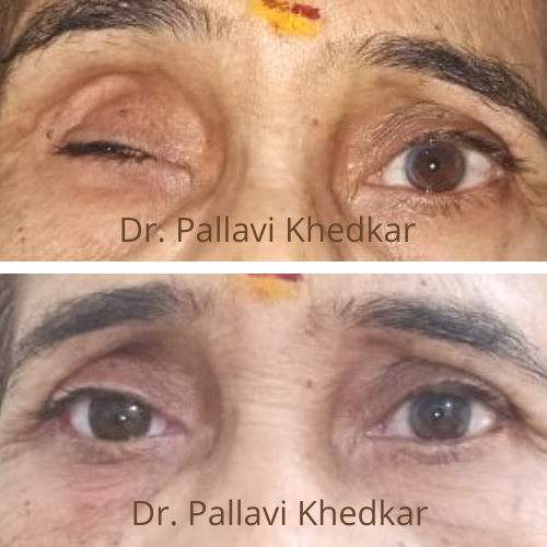
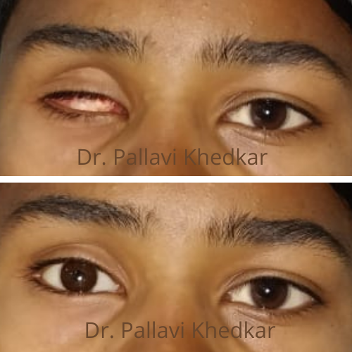
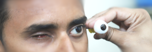
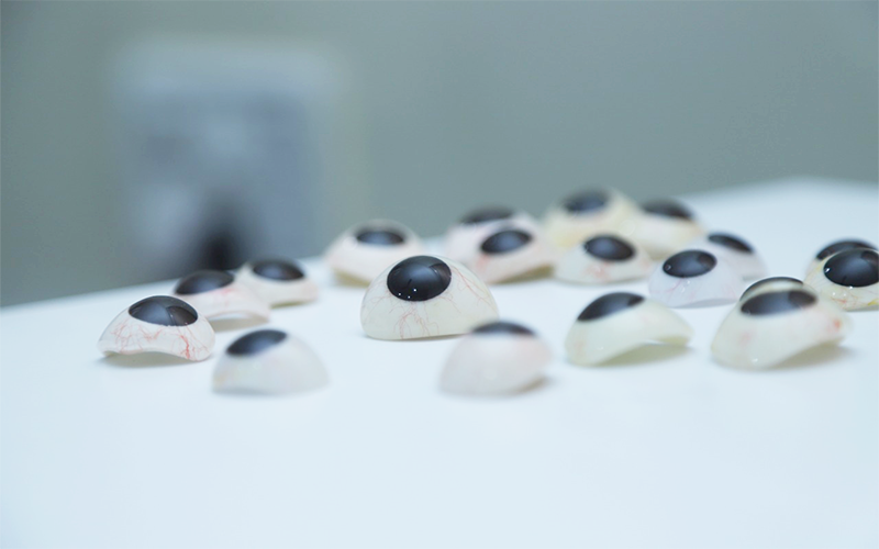

Treatment – Either an evisceration or an enucleation and place in the socket an orbital implant. Once the wound heals, an ocular prosthesis is placed over this. This surgery is performed by an oculoplastic surgeon. An ocular prosthesis is custom-built – to appear natural in size, shape, movement, and overall appearance. The entire process of creating an artificial eye or prosthesis is a painstakingly detailed one and needs specialized training and qualifications. Working along with an ocularist an outstanding customized ocular prosthesis is made. Some of the patients that Dr. Pallavi Khedkar and the ocularist have worked together on are shown here below.
The customized ocular prosthesis can be made at any age. The customized artificial eye is matched according to the opposite normal eye and can even be removed and repositioned by the patient.
Watering Eyes-Dacryology
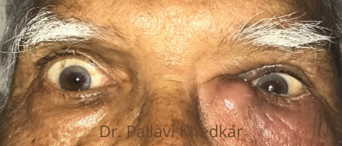
Blockage of the tear-draining duct causes infection of the lacrimal sac. When we blink, tears are forced towards the inner corner of the eyelid, where they drain via a punctum, a small hole. The tears then flow via the canaliculus, a very thin conduit, and into the lacrimal sac (near the inner corner of the eye). The tears then drain into the back of the nose and neck through a thin duct called the nasolacrimal duct, which is positioned inside the nasal cavity and drains into the back of the nose and throat.
Case 1
This is a case of 22 days old baby who presented with swelling near the eye at birth. Progressively it went on increasing over 20 days. He underwent nasal endoscopy and drainage of dacryocystocele endonasal under general anesthesia. His normal nasolacrimal passage was well maintained and patent after the procedure. The baby was relieved with symptoms immediately after the procedure. This procedure is done with a team of oculoplastic surgeon, ENT surgeon, and pediatric intensivist.
- Excessive watering.
- tears running down the cheek.
- mucus production.
- crusting of the eyelids is all signs of a tear duct obstruction.
- Dacryocystitis (infection and swelling of the lacrimal sac): Painful swelling and redness around the corner of the eyelid/nose junction. This illness has the potential to spread to the eyelids and possibly the face if left.
Topical and systemic antibiotics are used in the acute phase. The most common surgical procedure for a clogged tear duct is endoscopic DCR. In certain cases, a silicon stent is inserted during surgery and left in place for few weeks.
Droopy Eyelids/ Ptosis
Droopy Eyelids/ Ptosis
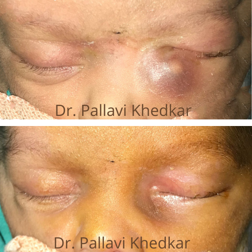
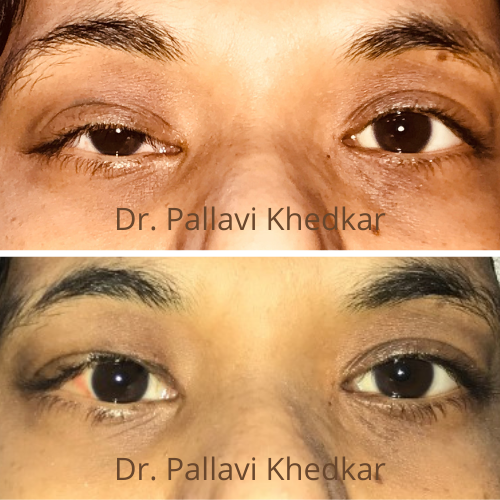
It is the medical term used for droopy eyelids. Ptosis (pronounced `toe-sis`) is a condition in which there is drooping of the upper eyelid. Ptosis occurs due to malfunctioning of the Levator palpebrae superioris (LPS) muscle, which is responsible for raising the upper eyelid.
- Congenital ptosis – Having uneven creases in the eyelids.
- Idiopathic.
- Post-traumatic.
- Neurological cause (Myasthenia Gravis).
- Tumors with nerve damage.
The primary symptom of ptosis is a visible drooping of the upper eyelid. Ptosis can cause a cosmetic disparity between the eyelids or the patient may have a ‘sleepy look’.
It may cause blurred vision or induce astigmatism
It can occur at any age
Ptosis in children needs urgent evaluation frequent raising of eyebrows and head tilting can indicate that ptosis is interfering with normal sight.
If the child’s eyelid droops so much that it interferes with the vision, amblyopia (lazy eye) can develop and that warrants early intervention.
Ptosis correction is a minor surgical procedure, involving the tightening of the muscle that lifts the eyelid. This can be performed through the eyelid skin or from the inside of the eyelid. The surgical approach taken depends on specific findings and testing performed during the pre-operative evaluation.
- Cosmetic concern
- Severe ptosis covering the pupil
- Head posture and amblyopia
Oculoplastic surgeons are the best specialists to treat this common problem as the surgery is a functional surgery and not purely a cosmetic one!
Surgical correction is the mainstay of treatment which provides a durable result if performed in time with the right principles.
Very few causes of ptosis are treated medically.
The surgical procedure may involve tightening the eyelid muscle or resection a part of the lax eyelid muscle. The purpose of the surgery is to match the positioning of both the eyelids and achieve symmetry.
Oculoplasty surgeons can decide about the method of treatment based on the strength of eyelid muscle and severity of ptosis.
Post-operative care
Usually, patients recover within 2 weeks after the surgical procedure
Mild pain and bruising may be noted, which is controlled by analgesics. Some patients may need a readjustment procedure after 7 to 10 days.
Facial Spasms
Have you ever noticed someone with a facial twitch that affects only half of their face? It looks as if they are about to sneeze, and one half of the face contorts suddenly? It’s almost as if it’s not in their control at all? And it keeps happening over and over again?
Hemi Facial Spasm
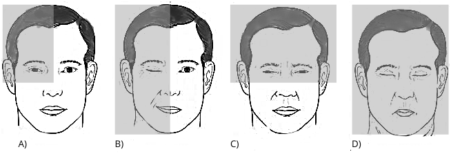
Hemi-facial spasm is involuntary twitching or contraction of most of the facial muscles on one side of the face.
All the muscles that twitch are innervated by the facial nerve. The facial nerve originates in the brain and travels across from behind the ear, in close relation to the salivary gland, and then supplies all the muscles of the face. This is the same nerve that helps you smile, pout, and whistle! But when inflamed, traumatized, under pressure, or damaged; this nerve can misfire and send out intermittent impulses which in turn causes involuntary spasms across one side of the face; right from the forehead right up to the neck muscles. Sometimes, a stroke or an infection may also cause it. However, in a substantial proportion of patients are often referred to as ‘idiopathic’ cases as there may not be any identifiable cause
Yes, there are different options available for the treatment of HFS. But before starting treatment, it is possible that your treating oculoplasty consultant may request a few investigations such as an MRI (Magnetic Resonance Imaging) and an EMG (electromyography)
Some medicines such as carbamazepine, benzodiazepines, and muscle relaxants may be used early on in the course of the disease or in situations when the patient refuses the option of botulinum toxin injection. Pharmacological therapy or medicines have not been as effective as Botulinum Toxin-A (Botox) in the resolution of symptoms.
What about surgery? In case there is documented compression over the nerve, then decompression surgery may be advised. Sometimes, some dilated blood vessels can cause a hemifacial spasm by compressing the facial nerve as it leaves the brainstem. Surgical decompression of these blood vessels may give good results.
Botox
Presenting symptoms:
- Frequent blinking
- Eyelids sometimes closed shut
- Eye irritation
- Sensitivity to bright lights
Treatment plan:
- BOTOX (Botulinum Toxin Type A) injections
- Artificial tears
- Dark sunglasses

Trauma Around Eye
Trauma Around Eye
Trauma to the face and area around the eye can occur in a road traffic accident, a direct hit, dog bite, or a blouse hook injury while breastfeeding.
Eyeball and surrounding soft tissue and a bony socket gets damaged.
Even though the trauma appears to be small, every eye injury should be treated by an ophthalmologist at the earliest.
Depending on the severity of the injury it is treated either conservatively or surgically.
Symptoms:
- Pain
- Decrease or loss of vision or double vision
- Tears on and around the eyelid
- One eye-popping out
- Blood in the clear part of the eye
- Unusual pupil size or shape
- Something embedded in or around the eye
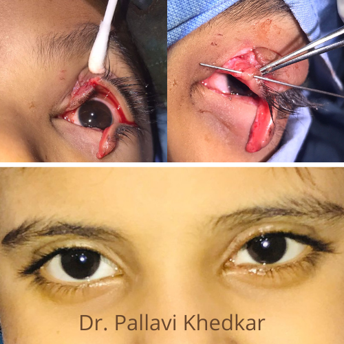
Any eye injury should be treated by a doctor, do not touch, rub, or attempt to remove any foreign body from the eye. Seek competent medical help right away.
Radiological imaging is needed in most cases of eye and orbital trauma.
The associated injuries like orbital fractures also need to be evaluated at the same time.
Eyelid tear with or without canalicular tear should be repaired at the earliest.
Eyelid trauma if repaired in time gives good cosmetic results.
Testimonials
मी कविता अंबेटकर कर सूचित करते की माझी मुलगी कादंबरी हि एप्रिल 2019 मध्ये घराच्या बाहेर खेळत असताना भटक्या कुत्र्यांनी तिच्यावर हल्ला करून तिच्या डोळ्याला व डोक्याभोवती गंभीर दुखापत केली होती. सदर दुखापत पाहिले असता आम्ही पूर्णतः घाबरून गेलो होतो व आता काय करायचे हा आमच्यापुढे फार मोठा प्रश्न निर्माण झाला होता. त्यानंतर मुलीला आम्ही अमृत बाल हॉस्पिटलमध्ये दाखल केले. तेथे आम्हाला तिच्या डोळ्याचे व पापण्यांचे ऑपरेशन करावे लागेल असे सांगण्यात आले.आशा वेळी काय निर्णय घ्यावा हे आम्हाला कळेनासे झाले. त्यावेळी संध्याकाळी डॉ. पल्लवी खेडकर यांची भेट झाली असता त्यांनी सर्व घटना समजून घेऊन दुखापत पाहिले असता, घाबरू नका यावर आपण योग्य तो निर्णय घेऊन तिचे ऑपरेशन व्यवस्थित पार पाडू असे आम्हाला सांगितले. त्यानंतर दुसऱ्या दिवशी डॉ. पल्लवी खेडकर यांनी अतिशय उत्कृष्टपणे ऑपरेशन करून तिची डोळ्याची झालेली गंभीर दुखापतीवर शस्त्रक्रिया केली. यानंतर आम्हाला खूप भीती अशी होती की मुलीची डोळ्याची गंभीर जखम पूर्ण होईल अथवा नाही. त्यानंतर आम्ही मॅडम कडे व्यवस्थितपणे ट्रीटमेंट घेतली आज माझ्या मुलीचा डोळा व झालेली गंभीर दुखापत पूर्णपणे बरी झालेली आहे. तिच्या डोळ्याकडे पाहिले असता तिला यापूर्वी जखम झाली होती असे मनात किंचितही वाटत नाही. त्यामुळे आज तिची दुखापत व्यवस्थित होण्याचे पूर्ण श्रेय डॉ. पल्लवी खेडकर यांनाच आहे. माझ्या कुटुंबाच्या वतीने डॉ. पल्लवी खेडकर यांचे शतशः आभार मानते. धन्यवाद
I am satisfied with the treatment and surgery by Dr. Pallavi, communication with the patient is very good. we are really satisfied. One of the best eye Hospitals. Would recommend it to anyone who has an eye problem, from the Doctor to staff all so professional and responds well with humility to your problem.Thank you.
अपघातात एक डोळा गमावल्या मुळे मला खूप दुःख झाले होते. डोळा गमावल्या पेक्षा आपला एक डोळा तिरकस ( तिरळे पण) दिसतो म्हणुन समाजात वावरतांना माझा आत्मविश्वास कमी झाला होता व नैराश्य आले होते.. परंतू गजानन हॉस्पिटल या ठिकाणी ऑक्युप्लास्टि शस्त्रक्रिया करण्यात आली म्हणून मी आज समाजात विश्वास पुर्वक वावरत आहे. व आता कुठल्याही प्रकारचा त्रास होत नाही. Thanks to Gajanan Hospital And Team.
Call Now For Appointment
"The eye is a great gift from the Lord. Take good care of it"
- Fill The Following Form

